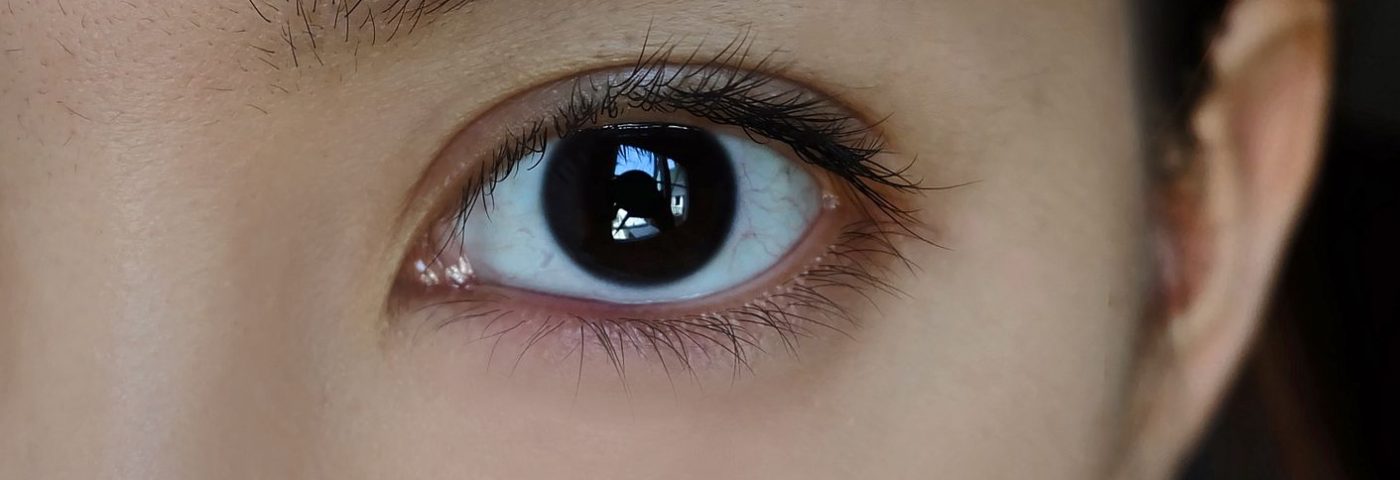
A case study described how a woman with a rare genetic autoimmune disorder developed severe neurotrophic keratitis (NK).
Researchers say the case highlights the importance of regular follow-ups and treatment of the corneal nerves in patients with an autoimmune disorder.
Titled “Neurotrophic keratitis in autoimmune polyglandular syndrome type 1: a case report,” the study was published in the journal BMC Ophthalmology.
Autoimmune polyglandular syndrome type 1, known as APS-1, is a genetic disease caused by mutations in the AIRE gene that lead to spontaneous autoimmunity, in which the immune system mistakenly attacks the body.
The rare disease is characterized by at least two of three factors that develop during childhood. Specifically, these are low levels of parathyroid hormone, known as hypoparathyroidism, primary adrenal insufficiency called Addison disease, and chronic fungal infections with Candida — a type of yeast — of the body cavities, skin, and nails. Such fungal infections are known as chronic mucocutaneous candidiasis.
Other symptoms include hair loss (alopecia), loss of skin pigment (vitiligo), and the wasting of dental enamel and nails. Eye damage also can occur, mostly reported as degeneration of the retina and keratoconjunctivitis with dry eye — simply put, inflammation of the tissue that lines the inside of the eyelids and covers the white of the eye.
Although damage to the cornea due to dry eye occurs in up to 50% of APS-1 patients, there have been no reports regarding the corneal nerve damage that underlies NK. There also has been no research investigating the relationship between APS-1 and NK.
Now, for the first time, researchers at the National Taiwan University Hospital, in Taiwan, have documented a case of a woman with APS-1 who was diagnosed with severe NK and ocular surface disease caused by dry eye.
The woman, age 27, was initially treated at the hospital for chronic blurred vision and discomfort. She had similar complaints 13 years before and was diagnosed with dry eye syndrome with corneal erosion in both eyes (keratopathy). While she had received topical treatment, it had led only to a partial recovery.
The patient had APS-1 with Addison disease as well as an autoimmune disorder involving chronic inflammation of the thyroid (Hashimoto thyroiditis), hypoparathyroidism, and hypogonadotropic hypogonadism, in which her ovaries produce little or no sex hormones.
She was short in height, at 147 centimeters (about 4 feet 8 inches tall), with low body weight — 37 kilograms or about 82 pounds — and had hair loss. While she had worn soft contact lenses for about five years, she had stopped wearing them three years previously.
An eye exam showed that her visual acuity was 20/40 in the right eye and 20/50 in the left eye, with two patches of corneal erosion in the right eye and superficial erosions on the lower corneal surface in the left eye. Of note, visual acuity measures an individual’s central vision, or the capacity to see detail, with a visual acuity of 20/20 referred to as normal vision; any score above 20/200 is considered legally blind.
Dry eye also was noted, along with meibomian gland deficiency, leading to a lack of oil that coats the eye surface to prevent dryness.
In vivo confocal microscopy (IVCM) of the eye found significant abnormalities of the corneal nerves, which is “not usually seen in patients with dry eye,” the researchers wrote.
Epithelial cells on the top layer of the cornea were elongated with a tendency to shed easily. The corneal nerve bundles were very thin and showed damage. As a result, ocular surface disease caused by dry eye with severe NK was diagnosed.
Corneal problems persisted in the following four months despite regular application of artificial tears, corticosteroids, the immunosuppressant cyclosporin, autologous serum eye drops, and nighttime ointment. Corneal erosions and inflammation were found in both eyes.
Treatment included therapeutic soft contact lenses and punctual occlusion, a dry eye procedure consists of inserting plugs into the tear drainage area to keep the tears in the eyes longer. Both treatments had a limited effect, and the patient continued to experience intermittent corneal erosions over the next two years.
To explain these new findings, the researchers suggested that the autoimmune imbalance in APS-1, or its hypothyroidism, may have directly caused damage to the corneal nerves.
Furthermore, studies have shown that a mouse model of AIRE gene deficiency — a model of autoimmunity — has a loss of nerves in the cornea, a lack of secretions, and dry eye. This supports the hypothesis that “APS-1 may inherently cause [nerve damage] and therefore lead to APS-1 keratopathy secondarily,” the researchers wrote.
“Patients with APS-1 may have ocular surface disease and severe damage to corneal nerves,” the investigators concluded. “Regular follow-up and treatment focusing on the regeneration of corneal nerves is particularly important in these patients.”

