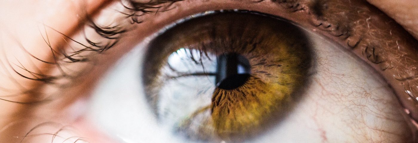
Two patients with herpes zoster ophthalmicus (HZO) developed an early and severe inflammation of both the cornea and the uveal tract — the pigmented inside lining of the eye — that manifested as neurotrophic keratitis, a case study reports.
The study, “Herpes Zoster Keratouveitis With Hypopyon and Hyphema” was published in the Taiwan Journal of Ophthalmology.
Herpes zoster ophthalmicus occurs following the reactivation of the varicella-zoster virus — the agent causing chickenpox and shingles — in the trigeminal nerve (the main nerve that serves the cornea, which is the transparent front layer of the eye).
Common ophtalmologic symptoms following HZO include blepharoconjuctivitis (an inflammation of the eyelids and conjunctiva), epithelial keratitis (an inflammation of the cornea surface), or anterior uveitis (an inflammation of the middle layer of the eye).
Of note, the conjuctiva is the thin layer that covers the white of the eye and the inner surface of the eyelids.
In this report, researchers at the University of Malaya Medical Centre in Kuala Lumpur, Malaysia, described the case of two women, ages 61 and 74, who presented with HZO complicated by early severe neurotrophic keratitis (NK).
The first patient had a two-day history of right eye redness and painful skin rashes on the right forehead and upper eyelid. Skin rashes were observed in the first division of the trigeminal nerve, but no evidence of Hutchinson’s sign was found. Hutchinson’s sign corresponds to vesicles on the nose tip, which occur before the development of ocular herpes zoster.
Further examination showed the patient had a swollen upper right eyelid and conjunctival injection, a mild generalized hemorrhage or a dilation of the conjuctival vessels that causes redness in the eye and commonly known as as bloodshot eyes. At this time, the right cornea and anterior chamber were clear. The anterior chamber is the front part of the eye between the cornea and the iris (the colored membrane of the eye).
The patient was treated with acyclovir, an anti-viral, both orally (800 milligrams) and topically, five times a day. Topical lubricants also were used.
After one week, her right eye’s vision declined, and Hutchinson’s sign was still evident. The patient also presented a large epithelial defect in the center of the eye and the anterior chamber was filled with hypopyon, a milky fluid that indicates infection, and hyphema, an accumulation of red blood cells. Corneal sensation was absent and intraocular pressure was above normal range.
Since these symptoms indicated a microbial infection, broad spectrum antibiotics were administered as eye drops together with antiglaucoma lubricants, cyclopedic (drugs to dilate the pupil) and acyclovir treatment was maintained. Microbial analysis of the cornea revealed an infection with Streptococcus pneumonia.
Due to the epithelial defect, two weeks into treatment a temporary surgical intervention called tarsorrhaphy, wherein eyelids are partially sewn together, was performed. This significantly helped resolve the microbial infection and the hypopyon and hyphema.
The patient’s pupil and lens were covered by a corneal edema (swelling), and the patient’s vision remained poor. By the third month cornea scarring and bleeding were observed.
The second HZO patient
The second patient presented with a three day history of blurry vision in the left eye, along with skin lesions affecting the area around the eye (called periorbital lesions) and eye redness for two weeks. Treatment with oral acyclovir and topical ciprofloxacin (an antibiotic) had been initiated the previous week.
Eye examination revealed the patient had an edema in the upper eyelid as well as the cornea. Similarly to the first patient, she also had hypopyon with hyphema. Corneal sensation was reduced, and, while negative at presentation, Hutchinson’s sign appeared the following day.
This patient was diagnosed with HZO with keratouvitis, which is inflammation of both the cornea and the uveal tract, the middle layer of the eye that includes the iris.
She underwent treatment with acyclovir eye ointment, lubricants, ciprofloxacin eyedrops and topical dexamethasone (a corticosteroid anti-inflammatory).
By day four, the epithelial defect persisted and a bandage contact lens was applied. This also significantly improved the hyphema and the hypopyon after one week. At this stage, intraocular pressure increased and anti-glaucoma medication was administered.
Although the epithelial defect was practically resolved with treatment, the patient presented with a recurrent defect at three months, and the corneal edema re-appeared together with hypopyon and hyphema.
Further treatment with dexamethasone (a steroid) reduced the hyphema, but no resolution of the cornea edema and epithelial defect were achieved. At the last follow-up, damage to the cornea persisted and eyesight was poor.
“Our cases describe an uncommon, early severe keratouveitis manifested as NK and hemorrhagic hypopyon following HZO in elderly immunocompetent individuals,” the researchers wrote.
According to the team, “an acute and severe manifestation of HZO occurring in the elderly population can present as NK in conjunction with hypopyon, hyphema, and skin rashes without Hutchinson’s sign.”
“An elderly patient when presenting to the clinician should be treated with the utmost vigilance as clinical sign, such as the Hutchinson’s sign, may be absent at its early phases. Nontraumatic hyphema and hypopyon associated with HZO as seen in these cases appear to be an early clinical sign leading to a poor visual outcome” they concluded.

